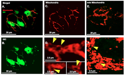

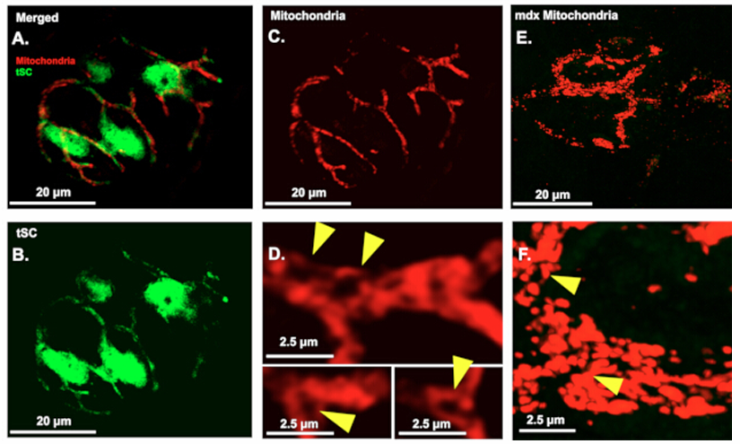
Under the microscope, mitochondria inside healthy terminal Schwann cells form a web of interconnected branches, with tiny doughnut-shaped loops appearing near where the cells branch and split.
But getting that view presents a challenge. Terminal Schwann cells — which help maintain and repair the connections between nerves and muscles — can begin changing shape the moment they are removed from their natural environment. That makes it difficult for scientists to study intracellular components as they would be in the body.
New research from Texas A&M University offers a way to get that closer look.
Published in the Journal of Visualized Experiments, the study outlines a method for imaging and evaluating mitochondria inside terminal Schwann cells while preserving their natural structure. This technique gives researchers a new tool for investigating how these cells respond to nerve injury, disease and recovery.
People are also reading…
“If you want to take a look at the things inside of them, as they are in the body, then somehow you have to image them without them knowing anything is disrupted,” said Dr. Aaron Morton, an assistant professor in Texas A&M’s Department of Kinesiology and Sport Management.
Cells that spring into action after injury
To explain the role of Schwann cells, Morton compares nerves to electrical wires.
A nerve is like the copper wire carrying the signal, while Schwann cells act like the plastic coating around it, providing insulation. But if the wire is damaged, the Schwann cells take on a different job.
After a nerve injury, Morton said, Schwann cells can change shape and move into the damaged area, where they help clear debris and release growth factors that support healing. The cells then “hold hands,” as Morton describes it, forming a pathway that guides the regenerating nerve.
Terminal Schwann cells are found at the neuromuscular junction — the point where a nerve communicates with a muscle. Normally, Morton said, they have a distinctive shape roughly resembling a pretzel. But when they sense injury or disruption, they can lose that structure, “turn into spheres” and begin moving around.
That ability to transform is important for recovery. But it also explains the challenge researchers face: Disturbing the cells to get a closer look can change the very structure they are trying to observe.
A window into mitochondrial health
Morton and his colleagues are particularly interested in the mitochondria inside these cells.
Best known as the “powerhouse of the cell,” mitochondria also form dynamic networks that connect and separate. Their shape and organization offer clues about how well they function and use oxygen to make energy. Given their function as oxygen consumers, this is also the first peer-reviewed report describing mitochondria as the “lungs of the cell,” which Morton suggests as a more effective way to describe them.
Studying that structure presents another problem. Terminal Schwann cells are embedded in muscle, which is packed with mitochondria of its own. A dye that labels mitochondria inside the Schwann cells can also light up those in the surrounding muscle.
The researchers developed a technique that labels mitochondria inside terminal Schwann cells before the dye spreads significantly into the surrounding muscle. Researchers then quickly prepare the tissue for imaging to minimize changes to the cells.
To demonstrate what the method could reveal, researchers applied it in healthy and dystrophic tissues.
In healthy terminal Schwann cells, mitochondria formed interconnected networks with small, doughnut-shaped loops. In the dystrophic model, those networks were more fragmented and had lost those configurations.
“When everyone’s happy, they hold hands and exchange contents,” Morton said. “When they’re mad, they all break apart and keep to themselves.”
The comparison showed that the technique could capture differences in mitochondrial organization between healthy and dystrophic terminal Schwann cells.
Opening the door to new questions
That ability could help address a larger gap in scientists’ understanding of terminal Schwann cells.
Research on terminal Schwann cells has lagged behind that of many other cell types, Morton said, in part because scientists have not had the tools to investigate every aspect of them in their natural environment.
One potential application is studying neuromotor disease. Previous research has implicated terminal Schwann cells in amyotrophic lateral sclerosis (ALS), diabetic neuropathy and spinal muscular atrophy. The current study did not investigate those diseases, but the new method could give researchers another way to study what happens inside these cells.
Morton’s lab also plans to use the technique to study nerve injury, including whether an experimental biomaterial that promotes mitochondrial health could improve nerve recovery.
For Morton, that is the value of developing a new way to see these cells: It creates opportunities to ask questions researchers could not answer before.
“When a tool is created or when a new method is made, oftentimes it opens the door to new investigations, opening the door to new answers,” Morton said.